

(A downloadable PDF of this article is available at the end. The PDF includes the original images and formatting.)
Drug and diagnostic development rely heavily on models. When these models lack biological context or miss important features of human disease, promising laboratory results often become costly clinical failures or contribute to persistent challenges in diagnostic performance. Primary human tissue linked to longitudinal clinical data addresses many of these gaps by connecting molecular signals with disease biology and patient context.
Most early-stage development uses immortalized cell lines. Since the 1950s, these cells have contributed enormously to biomedical research as they have become widely available, inexpensive, reproducible, scalable, and indispensable for certain kinds of experiments.
Immortalized cell lines are well adapted for laboratory growth, but this adaptation has a drawback: it creates biological distance from the original patient tissue. During prolonged culture, cells undergo selective pressure, leading to genetic and epigenetic changes that favor laboratory propagation rather than native function. As these adaptations accumulate, the high-resolution biological data required to understand and solve complex drug and diagnostic problems is lost.
For early mechanistic work and high-throughput screening, cell lines are often sufficient. In high-cost, high-stakes scenarios where intricate details about cellular heterogeneity and patient-specific biology will ultimately determine success or failure, direct human biological evidence is essential. This evidence can be obtained only through direct access to the patient or by analyzing primary tissue linked to relevant clinical data.
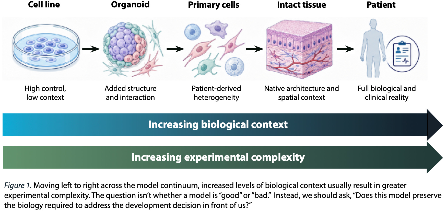
Primary tissue and patient-derived cells provide higher-grade evidence because they retain detailed biological variation, including the following:
Cellular heterogeneity. Primary tissue allows us to see complex disease microenvironments such as diverse immune or malignant subpopulations, therapy-resistant subclones, and stem-like cells that drive relapse and progression; in cell lines, these subpopulations are compressed into more uniform populations, obscuring unique responses to therapy and cellular interactions.
Patient-specific context. Primary tissue provides context across different patients, allows us to identify patient subgroups, and helps us optimize biomarker strategies to lessen the risk of target failure in heterogeneous populations; cell lines capture a single biological history and do not represent variation across populations.
Tissue architecture. Primary tissue preserves critical spatial context, such as tumor-immune cell interactions, which determine target localization and response potential; over time, cell lines alter gene expression and silence tissue- specific functions, making validation in human specimens essential to confirm findings.
Longitudinal clinical data. Linking primary specimens to longitudinal clinical records places molecular findings in clinical context, allowing us to assess how patient genotypes, prior therapies, and variations in disease stage might be influencing molecular signaling and therapeutic outcomes; cell lines provide little or no relevant data about an individual’s clinical history or disease progression.
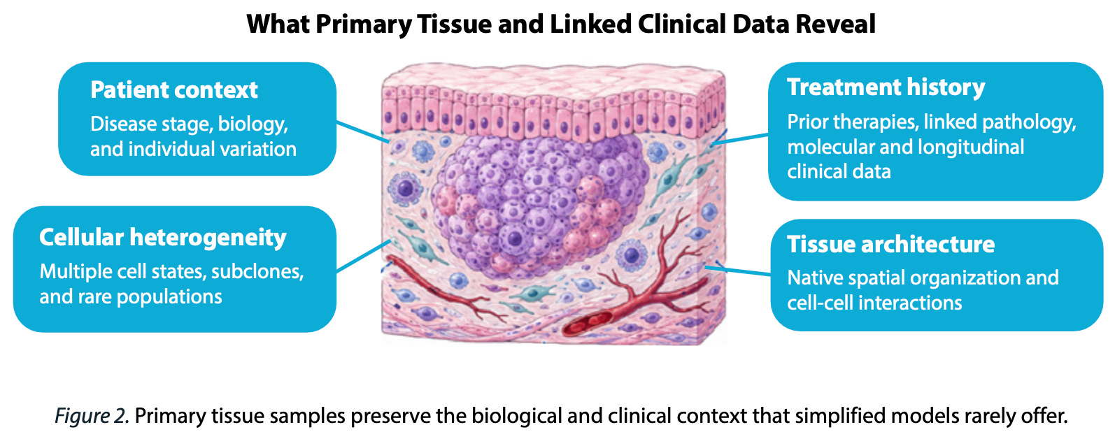
In immune-mediated diseases like rheumatoid arthritis, direct study of affected tissue can identify the interacting fibroblast, myeloid, B-cell, and T-cell populations that maintain inflammation and distinguish biologically different forms of disease that may require different therapeutic strategies.
In oncology, primary specimens can reveal whether a therapeutic target is expressed on the cells that drive disease, whether that expression is consistent across patients, whether normal cells share the same target, and how different malignant and immune populations respond to treatment.
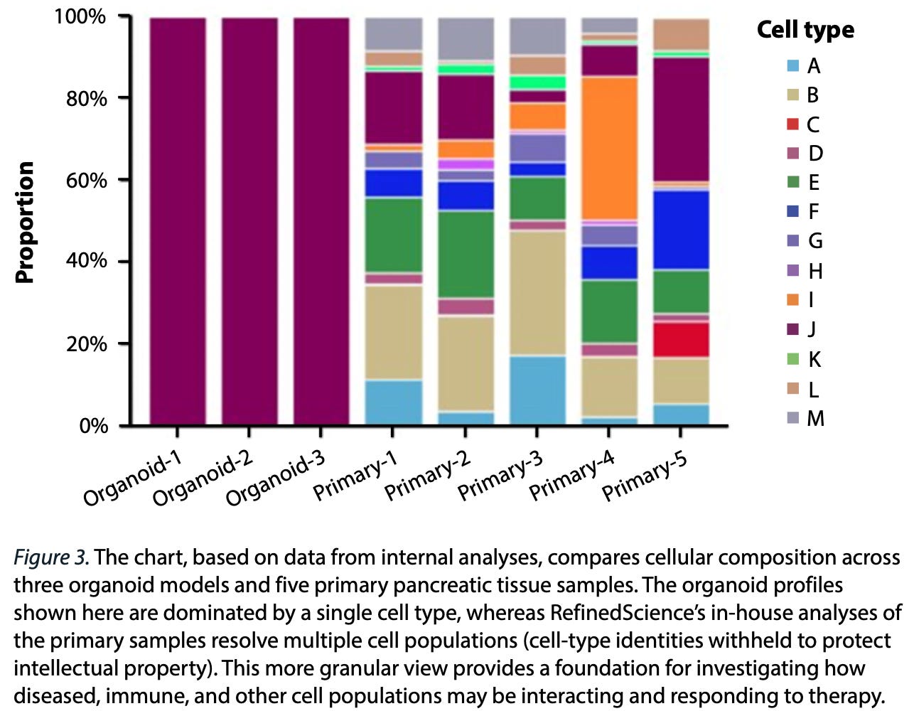
Combining this deeper level of cellular detail and molecular diversity with linked clinical data and de-identified patient histories allows us to generate intellectual property and provide our partners with extensive disease insights they would not get from cell lines or other conventional methods.
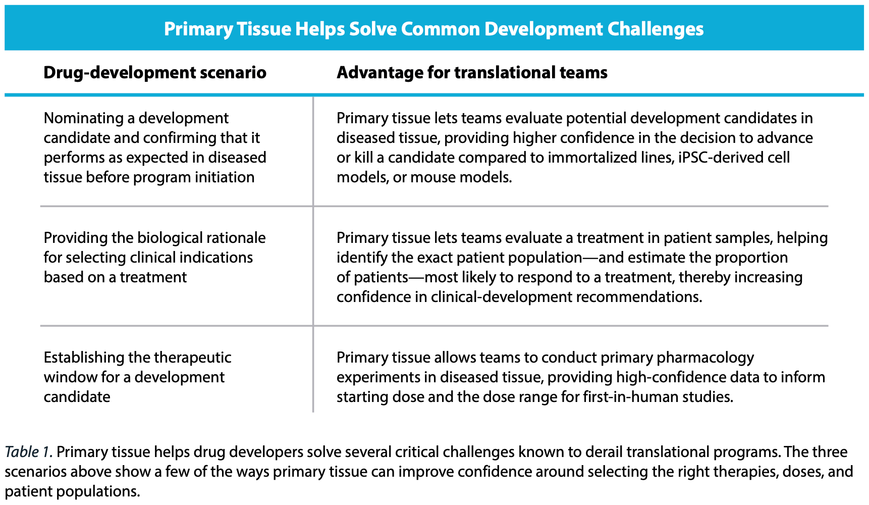
Because of these advantages, primary human tissue should occupy a more central position in translational research. By integrating well-characterized human tissue with clinical records, single-cell profiling, and functional experimentation, our approach bridges the gap between low-resolution modeling and actual human biology, ensuring therapies and diagnostics are grounded in the complex, native environments they will ultimately encounter.
Primary tissue and the next generation of biological models
Generative AI models are creating new opportunities to predict cellular state, drug response, target relationships, and outcomes of perturbation experiments. But, as stated earlier, a model learns from the biological contexts represented in its training data. A model trained predominantly on a limited set of immortalized systems may learn responses that are internally consistent but fail when applied to primary specimens with variation among cell types and differences in disease stage, co-mutations, tissue environments, treatment exposures, and patient-outcome histories.
The central challenge in training better AI models is not obtaining more cells, but capturing more relevant biological contexts. In this framework, the durable asset is the algorithm plus the combination of well-characterized tissue, linked clinical information, high-quality molecular data, functional experimentation, and consistent data-generation practices—all of which contribute to a positive feedback loop that can improve model integrity while generating novel questions, hypotheses, and findings.
Biological models trained on primary-tissue data rather than data derived solely from cell lines and other conventional systems will likely become the best-performing, most sought-after AI models in biotechnology. These will be the models that help move generative AI from a discovery tool to a true end-to-end tool for drug and diagnostic development.
Download the report
Prefer to save or print a copy? Download the complete PDF below, including the images and formatting.

